Lay-Language Papers
Multiplexing Radiography for Ultra-Fast Computed Tomography
J. Zhang, G. Yang, Y. Lee, S. Chang, J. Lu, O. Zhou
University of North Carolina at Chapel Hill
jzh@physics.unc.edu
Author description of paper MO-E-L100J-7
Monday, July 23, 2007
2007 AAPM Meeting, Minneapolis
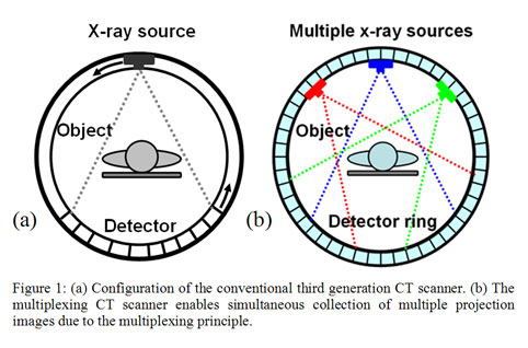
The development of computed tomography (CT) technology is one of the most important breakthroughs in the field of imaging. CT scanners are now widely used for diagnostic medical imaging and security screening. In today’s state-of-the-art medical and security CT scanners, over 1,000 images are collected in less than one second by high-speed rotation of an x-ray tube around the object, as illustrated in Figure 1a. However, the data is collected in "serial" fashion, or essentially one piece of data at a time. The inefficient serial data collection scheme severely limits data collection speed, which is critical for imaging of objects in rapid motion such as for diagnosing cardiovascular diseases, acquiring high-quality images of tumors inside patients, and inspecting airport luggage. Further increasing CT speed demands increasing the peak power of x-ray sources and increasing the rotation speed of the gantry, the device that revolves the x-rays around the object to be imaged. For both of these variables, these technologies have approached their physical limits. To illustrate how far current devices have been pushed, the large x-ray tubes in modern CT scanners are subject to an acceleration force larger than 30 G during scanning.
Multiplexing represents an innovative solution for potentially speeding up CT scans. A widely used concept in many communications-related fields, multiplexing is a process of combining multiple signals to form one composite signal for transmission. It's analogous having a multilane highway to accommodate a large volume of cars, then allowing them eventually to exit in a single-lane ramp. Serial transmission, however, is analogous to having just a single-lane road at all times for all vehicles.
Since multiplexing involves transmitting many signals simultaneously, the approach holds the promise of significantly increasing data transmission rates. Multiplexing has been studied and used in applications such as high-speed computer modems, cellular phones and high-density magnetic and optical recording. However, it has not been applied to x-ray radiography, mainly due to limitations of the current technology of x-ray sources. Difficulty in modulating, or rapidly changing, the properties of x-rays is one of these limitations, as the metal filament-based x-ray tubes need warm-up or cool-down time. Another limitation comes from most x-ray tubes being single-pixel devices, only capable of generating one x-ray beam at a time.
Electron field emission technology can mitigate the limitations of the current x-ray sources. In a conventional x-ray tube, the metal filament generates electrons when exposed to very high heat, around 1,000 C. Those electrons are bounced off a separate piece of metal, generating x-rays. Carbon nanotubes, on the other hand, emit electrons at room temperature under an externally applied electric field, which is an instantaneous process. And also because there is no heat, the nanotube x-ray machine is much smaller, as it is free of bulky insulation. The smaller size makes it possible to integrate multiple x-ray sources together to form a multi-pixel x-ray source.
We have been developing the possibilities of multiplexed CT imaging. Recent experiments using carbon nanotube-based field emission devices have demonstrated the capability of generating x-rays with the desired properties for multiplexed transmission. In 2005, we developed a prototype CT scanner with multiple x-ray sources, called a multi-pixel scanner. The machine required no mechanical motion but switched rapidly among many x-ray sources, each taking an image of the object from a different angle in fast succession. Very recently we combined this multiple x-ray-source innovation with multiplexing techniques to demonstrate a new CT scanning system, called a multiplexing CT scanner, as shown in Figure 1b. We recently created a 25-pixel multiplexing CT scanner, but engineering difficulties lie in front of the ultimate goal, a scanner with approximately 1,000 x-ray pixels. The cost of these machines would not rise significantly, as carbon nanotube field emission x-ray technology enables hundreds of x-ray cathodes to be fabricated on a single silicon wafer.
For the multiplexing CT scanner, all the x-ray sources are turned on simultaneously to capture images from multiple views at the same time. Each x-ray signal is modulated at a unique frequency, similar to how a radio station transmits its audio signal by modulating the signal at a unique frequency that corresponds to the station's number on the dial. Together, the multiple x-ray signals form a composite signal for transmission. The composite signal is captured by the x-ray detector, and then is separated into individual signals, just as a radio receiver separates dozens of signals coming from multiple stations into individual locations on the dial. The multiplexing CT scanner has the potential to significantly increase the imaging speed for CT scanning without compromising the imaging quality. In general a factor of N/2 (N=total number of images) increase in the speed can be achieved using the multiplexing technique. For example, the speed of clinical CT scanners that can acquire around 1,000 views per gantry rotation would increase by a factor of 500.