Single-Topic News Release
256-Slice CT Scanner Promises Faster Heart Images, Lower Radiation Doses
At the annual meeting of the American Association of Physicists in Medicine in Orlando on July 31, Richard T. Mather, Ph.D. of Toshiba America Medical Systems (rmather@tams.com) in Tustin, California will describe a work-in-progress (WIP) prototype of the wide-area-detector "256-slice" CT scanner. 256-slice CT is a new imaging technology that can provide detailed images of the entire heart, including the coronary arteries, in less than half a second while requiring a fraction of the radiation dose that is traditionally needed [potentially as low as 2 milliSieverts (mSv) as opposed to the 13 mSv with present state of the art CT equipment].
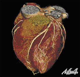
This image of the heart was acquired by a "64-slice" CT scanner, the current state-of-the-art in clinical CT imaging, as it rotated an x-ray beam several times around a patient. A prototype 256-slice CT scanner, slated to be introduced clinically within the next few years, can image the entire heart in a single rotation around the patient, providing faster scans with potentially lower radiation doses. (Image courtesy Toshiba America Medical Systems)
A followup to clinically successful "64 slice" CT machines, the 256-slice scanner, slated to be introduced clinically within the next 2-3 years, has the potential to routinely detect coronary artery disease and heart defects in a single test during an emergency-room visit or annual physical. For this reason, Mather says, he and his colleagues hope this technology can help reduce healthcare costs, by potentially eliminating batteries of unneeded tests in certain cases.
By rotating a beam of x-rays around a patient, computed tomography (CT) machines obtain detailed cross-sectional images of body organs, such as the heart. To create the cross-sections, CT detectors split up the organ into various pieces or "slices," with a higher number of slices delivering more detail (or alternately, enabling the scanner to image a larger region of the body during a single pass). At the same time, having more slices reduces the amount of time and can reduce the radiation required to create the images. 64-slice CT machines, with their ability to cover 32 mm of body anatomy and provide slices just 0.5 mm in size, can provide high-detail pictures of the entire tree of coronary arteries in 6-9 seconds. By comparison, the 256-slice design can image 128mm at a time, covering the entire heart, with the same level of detail as the 64-slice CT, in an even shorter time, over a single rotation of the scanner, or less than 500 milliseconds.
When used in conjunction with an injected dye (contrast agent) that highlights the blood vessels in the heart, the rapid single-rotation coverage of the machine enables it to obtain valuable information on blood flow in the heart, and potentially uncover heart defects such as coronary stenosis, narrowing of the heart’s blood vessels, and cardiac shunts, the undesirable mixing of blood from the different heart chambers.
One of the main differences between Toshiba’s 256-slice system and today’s premium commercial scanners is that the 256 does not require the scanner to move the x-rays in a helix-shaped trajectory to image the brain, heart, and other major organs. By eliminating the need for such helical scanning, and instead acquiring the images without moving the patient, the new design brings an inherent improvement in image quality and the significant reduction in patient dose.
A challenge in developing the 256-slice CT scanner, Mather says, is that it forces advances in accompanying technologies. In addition to pushing forward the performance of wide-area detectors, the researchers are addressing existing limitations in image reconstruction, image processing, and data transfer technology as they prepare to bring the device to market.
Meeting Paper: Monday, MO-E-330D-2, "256 Slice CT: Development, Design, and Clinical Applications", July 31, 2006, 4:25PM, Room 330 D. Click Here for Technical Abstract
Presented at: 48th Annual Meeting of the American Association of Physicists in Medicine, July 30-August 3, 2006, Orange County Convention Center, Orlando, FL. Click Here for Meeting Homepage
ABOUT AAPM
AAPM (www.aapm.org) is a scientific, educational, and professional organization of more than 6,000 medical physicists. Headquarters are located at the American Center for Physics in College Park, MD. Publications include a scientific journal ("Medical Physics"), technical reports, and symposium proceedings.
###
For more information, please contact Ben Stein of the American Institute of Physics, bstein@aip.org, 301-209-3091, Jeff Limmer, AAPM Media Relations Subcommittee Chair, jeffl@aspirus.org, or Martha Heil, American Institute of Physics, 301-209-3088, mheil@aip.org