

Virtual Press Room
Press Conference
A PRESS CONFERENCE presenting three important studies from the AAPM Annual Meeting will be held on Wednesday, July 15, 10:00 a.m. – 11 a.m. PT, in Room 208B, Level 2 of the Anaheim Convention Center.
The press conference will include research on:

- Mammography risks overestimated: Large study shows women receive about 30 percent less radiation than previously believed.
- Common side effect of prostate cancer treatment can be reduced: A study of 1,001 prostate cancer patients suggests that factoring in anatomy and the radiation therapy dose can help doctors predict a patient’s risk of suffering from rectal bleeding, and tailor treatment to prevent the problem.
- Fetal radiation dose: Medical physicists will present research on a new technique to more accurately calculate radiation dose to a fetus.
For more information on the press conference, including teleconferencing details for those unable to attend in person, please contact: Matt Sobczak at 312-558-1770 or msobczak@pcipr.com.
Press Releases
Embargoed until Wednesday, July 15, at 10:00 a.m. (US Pacific Time). Please check back after that time.
- Risks Of Screening Mammography Overestimated
- Common Side Effect Of Radiation Treatment For Prostate Cancer, Can Be Reduced By Factoring In Anatomy, Therapy Dose
- Simple Formula Can Determine Radiation Dose to Fetus in Pregnant Women Who Have CT Scan
For detailed author information, contact: Matt Sobczak at 312-558-1770 or msobczak@pcipr.com.
Hot Topics
The 57th AAPM Annual Meeting features cutting edge research across the field of medical physics science. Four HOT TOPICS from the Scientific Program are summarized below. All hot topics are embargoed until the day and time of their presentation (noted in red).
Embargoed for Release until 1:45 p.m. PT July 15, 2015
Mobile CT Scanner Could Help Diagnose Brain Injury on the Spot
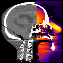 A mobile computed tomography (CT) scanner – significantly smaller than standard machines – may one day help doctors diagnose traumatic brain injury in patients on site, from football stadiums to the frontlines of combat. CT is an excellent method for detection of traumatic brain injury such as bleeding in the brain, but standard CT scanners are quite large, so patients must be brought to the machine to undergo testing, which delays the potential diagnosis. Researchers at Johns Hopkins University developed a prototype mobile scanner based on improved, high-quality-image cone-beam CT that could overcome logistical issues and be used to quickly assess whether a patient has suffered a traumatic brain injury. Among the many potential uses: in the intensive care unit (for bed-ridden patients who cannot risk transfer to a CT scanner), at urgent care and concussion clinics, in ambulance and mobile clinics, at stadiums and other athletic venues and in the military theater.
Speakers:
J.H. Siewerdsen, Ph.D., Professor, Johns Hopkins University, Baltimore
J. Xu, Graduate student, Johns Hopkins University
A. Sisniega, Ph.D., Graduate student, Johns Hopkins University
W Zbijewski, Ph.D., Research Associate, Johns Hopkins University
H. Dang, Graduate student, Johns Hopkins University
J.W. Stayman, Ph.D., Assistant Professor, Johns Hopkins University
X. Wang, Ph.D., Research Scientist, Carestream Health, Rochester, N.Y.
D.H. Foos, Ph.D., Research Scientist, Carestream Health
N. Aygun, M.D., Assistant Professor, Johns Hopkins University
V. Koliatsos, M.D., Assistant Professor, Johns Hopkins University
Additional studies related to this topic »
WE-EF-207-3 |
Design and Optimization of a CBCT Head Scanner for Detection of Acute Intracranial Hemorrhage J. Xu*, A Sisniega, W. Zbijewski, H. Dang, J. Stayman, X. Wang, D. Foos, N. Aygun, V. Koliatsos, J. Siewerdsen |
| WE-EF-207-5 | Monte Carlo Dosimetry for a Dedicated Cone-Beam CT Head Scanner A. Sisniega*, W. Zbijewski, J. Xu, H. Dang, J. Stayman, N. Aygun, V. Koliatsos, X. Wang, D. Foos, J. Siewerdsen |
Embargoed for Release until 7:30 a.m. PT July 16, 2015
Thermal Brachytherapy May Improve Treatment of Prostate Cancer
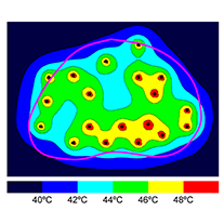 An innovative technology is being developed at the University of Toledo Medical Center involving radioactive seeds (brachytherapy) that can simultaneously deliver hyperthermia (extreme temperatures) to cancerous tissue to boost the effectiveness of radiation dose, more effectively killing cancer cells with minimal harm to surrounding healthy tissue. Traditional brachytherapy involves implanting small radioactive seeds into a tumor, where they emit radiation to kill the cancer cells. This recent innovation produces both radiation and heat from the same implanted seeds. The researchers created computer models to determine that their new thermobrachy seeds can be injected into prostate tumors and bring them to hyperthermia-sufficient temperatures when placed in a magnetic field. This new seed also has a self-regulating capability, able to plateau at the desired temperature and effectively treat the affected tissue, eliminating the need for invasive thermometry. The combination of hyperthermia and radiation can help improve oncologists’ ability to manage prostate cancer without increasing the dose of radiation, which can damage healthy tissue.
Speakers:
E. Ishmael Parsai, Ph.D., Professor and Director of Medical Physics, University of Toledo, Ohio Medical Center
D. Shvydka, Ph.D., Assistant Professor, Department of Radiation Oncology, University of Toledo
G. Warrell, Ph.D. candidate in Medical Physics, University of Toledo
Additional studies related to this topic »
| TU-AB-BRA-4 | Quantitative Radiomics: Sensitivity of PET Textural Features to Image Acquisition and Reconstruction Parameters Implies the Need for Standards M. Nyflot*, F. Yang, D. Byrd, S. Bowen, G. Sandison, P. Kinahan |
| TU-AB-BRA-7 | Radiomics of Breast Cancer: A Robustness Study N. Antropova*, M. Giger, H. Li, K. Drukker, L. Lan |
| TU-AB-BRA-8 | Radiomics in the Analysis of Breast Cancer Heterogeneity On DCE-MRI H. Li*, L. Lan, K. Drukker, C. Perou, M. Giger |
| TU-AB-BRA-9 | Radiomics and Radiogenomics for Breast Cancer Using Magnetic Resonance Imaging J. Oh*, E. Sutton, H. Veeraraghavan, A. Apte, E. Morris, J. Deasy |
Embargoed for Release until 10:15 a.m. PT July 14, 2015
Big Data Can Personalize Lung Cancer Therapy
 Combining non-invasive radiomics with an individual’s genetic makeup could help doctors provide personalized treatment to cancer patients. Researchers from five institutions analyzed computed tomography (CT) scans, gene-expression profiles, and outcomes of 351 lung cancer patients treated at two institutions and identified factors that doctors can use to predict how a patient’s tumor would react to treatment without having to undergo an invasive biopsy. This integrative big data approach could be used to determine which type of treatment would be the most beneficial for that patient, instead of relying on standard therapy without utilizing this information. This personalized medical care would help people receive therapy that is more likely to be effective in a timely and cost-effective manner.
Speakers:
P. Grossmann, M.Sc., Ph.D. student, Dana-Farber Cancer Institute/Harvard Medical School, Boston
O. Grove, Ph.D., Research Scientist, H. Lee Moffitt Cancer Center and Research Institute, Tampa, Fl.
N. El-Hachem, M.Sc., Ph.D. student, Institut de Recherches Cliniques de Montreal
E. Rios Velazquez, Ph.D., Post-doc, Dana-Farber Cancer Institute/Harvard Medical School
C. Parmer, M.Sc., Ph.D. student, Dana-Farber Cancer Institute/Harvard Medical School
R. Leijenaar, M.Sc., Ph.D. student, Research Institute GROW, Maastricht, Netherlands
B. Haibe-Kains, Ph.D., Assistant Professor, Princess Margaret Cancer Centre, Toronto
P. Lambin, M.D., Ph.D., Professor, Research Institute GROW
R. Gilles, Ph.D., Professor, H. Lee Moffitt Cancer Center and Research Institute
H. Aerts, Ph.D., Assistant Professor, Dana-Farber Cancer Institute/Harvard Medical School
Additional studies related to this topic »
Embargoed for Release until 10:15 a.m. PT July 14, 2015
Helping Doctors Adjust Lung Cancer Treatment to Reduce Lung Inflammation
 A study of 198 lung cancer patients treated with radiation therapy suggests big data information can be used to help doctors determine which patients are most likely to suffer from a common side effect of the treatment, pneumonitis, or inflammation of the lungs. Patients who suffer from this side effect often have shortness of breath, difficulty breathing during activity and coughing. Researchers collected a total of 3,888 “features” based on CT images, personal information (such as smoking status) and radiation doses, and created a framework for predicting a patient’s likelihood of developing pneumonitis. If validated, doctors could use this information to assess each patient and adjust treatment to decrease the risk of pneumonitis.
Speakers:
S. Krafft, B.S., P.S.M. Graduate Research Assistant, The University of Texas MD Anderson Cancer Center, Houston, The University of Texas Graduate School of Biomedical Sciences, Houston
T Briere, Ph.D., Assistant Professor, The University of Texas MD Anderson Cancer Center
L. Court, Ph.D., Assistant Professor, The University of Texas MD Anderson Cancer Center
M. Martel, Ph.D., Professor, The University of Texas MD Anderson Cancer Center
Additional studies related to this topic »
Featured Interviews
AAPM: Future Trends in Medical Physics
American Association of Physicists in Medicine (AAPM) President John Boone, PhD, discusses the impact of imaging in medical physics, as well as key topics addressed at this year’s meeting, with Imaging Technology News Editorial Director Melinda Taschetta-Millane.
AAPM: Size-Specific, Scanner-Independent Fetal Dose Estimates in Abdominal and Pelvic CT Examinations of Pregnant Patients
During the American Association of Physicists in Medicine (AAPM) 57th Annual Meeting in Anaheim, California, Imaging Technology News’ editorial director Melinda Taschetta-Millane spoke with Michael F. McNitt-Gray, PhD, FAAPM, about his abstract on Size-Specific, Scanner-Independent Fetal Dose Estimates in Abdominal and Pelvic CT Examinations of Pregnant Patients, which was presented at the meeting.
AAPM: Monte Carlo Simulation of Glandular Breast Dose in Mammography Using Breast CT-Derived Glandular Distributions
During the American Association of Physicists in Medicine (AAPM) 57th Annual Meeting in Anaheim, California, graduate student researcher at the University of California/Davis Andrew M. Hernandez discussed screening and dose distribution for women with dense breasts, and its implications for mammography.
AAPM: Dose-Volume Relations for Late Rectal Bleeding in 1001 Patients From Five Prostate Cancer Cohorts
Joseph Deasy, PhD, discusses is study on dose-volume relations for late rectal bleeding in 1001 patients from five prostate cancer cohorts with Imaging Technology News Editorial Director Melinda Taschetta-Millane.
AAPM gratefully acknowledges the cooperation of ITN in conducting these interviews and making them available here and on their website.